Human Airway Analysis
Jaesung Lee and Shuang Liu
 Computed tomography (CT) is widely performed for patients with chronic
obstructive pulmonary disease (COPD) and the airway dimensions measured
from CT chest scans have been demonstrated repeatedly to be correlated with
measures of airflow obstruction. Airway dimensions and characteristics vary
significantly for different regions of lung and for different anatomical or
bifurcation-based generations, therefore performing reproducible and
comparable measurements in airway requires the ability to locate the
corresponding positions to measure in different scans, which is challenging in
fully automated studies.
The purpose of this study was to develop a fully automated framework that allows
reproducible and comparable quantification of airway dimensions from low dose
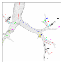
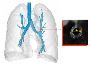
chest CT scans. The human airway is first segmented and labeled with anatomical
branch names. Then the lumen diameter and wall thickness are measured at each
anatomical branches and further averaged over each anatomical generations.
The presented framework was applied to 2360 low-dose chest CT scans and obtained
correct anatomical labeling in 90% (2124 out of 2360) of the scans based on
subjective visual evaluation.
 The Airway dimension measurements were validated using 14 CT scan pairs [2].
The lumen diameter can be measured within the precision of +/-0.8mm,
and the wall thickness can be measured within the precision of +/-0.5mm.
The precision in measuring overall airway dimensions within individual patients
was also evaluated [4]. The overall measures in individual patients the
lumen diameter can be measured precisely within +/-0.5mm , and the wall
thickness can be measured mmwithin +/-0.15mm.
Presentations and Publications
- J. Lee, A. P. Reeves, D. F. Yankelevitz, and C. I. Henschke.
Bronchial Segment Matching in Low-dose Lung CT Scan Pairs.
Proceedings of SPIE Medical Imaging,7660:72600A, Feb 2009.
- J. Lee, A. P. Reeves, D. F. Yankelevitz, and C. I. Henschke.
Assessment of Airway Dimensions in Low-dose CT.
RSNA 94th Scientific Assembly and Annual Meeting, Nov 2008.
- J. Lee, A. P. Reeves, D. F. Yankelevitz, and C. I. Henschke.
Skewness Reduction Approach for Measuring Airway Wall Thickness.
Int J CARS, vol.3, pp. S50-S52, June 2008.
- J. Lee, A. P. Reeves, S. Fotin, T. Apanasovich, and D. F. Yankelevitz.
Human Airway Measurement from CT Images.
Proceedings of SPIE Medical Imaging, 6915:691518, Feb 2008.
- J. Lee, A. P. Reeves, S. Fotin, T. Apanasovich, D. F. Yankelevitz,
and C. I. Henschke. Precision of Automated Airway Measurements in CT Images.
RSNA 93rd Scientific Assembly and Annual Meeting, Nov. 2007.
List of Current Research Projects
|

 Computed tomography (CT) is widely performed for patients with chronic
obstructive pulmonary disease (COPD) and the airway dimensions measured
from CT chest scans have been demonstrated repeatedly to be correlated with
measures of airflow obstruction. Airway dimensions and characteristics vary
significantly for different regions of lung and for different anatomical or
bifurcation-based generations, therefore performing reproducible and
comparable measurements in airway requires the ability to locate the
corresponding positions to measure in different scans, which is challenging in
fully automated studies.
 The Airway dimension measurements were validated using 14 CT scan pairs [2].
The lumen diameter can be measured within the precision of +/-0.8mm,
and the wall thickness can be measured within the precision of +/-0.5mm.
The precision in measuring overall airway dimensions within individual patients
was also evaluated [4]. The overall measures in individual patients the
lumen diameter can be measured precisely within +/-0.5mm , and the wall
thickness can be measured mmwithin +/-0.15mm.